A process to know and study chromosomes known as karyotyping. To do so, we need to culture and harvest chromosomes. Also, the present cytogenetic technique is very much tedious and time-consuming. We need a high level of experience and expertise to achieve results.
But don’t worry, It is possible to even without going to a wet lab. Trust me this blog is all about karyotyping and related activities. Here in the present article, I will explain to you some of the examples of karyotyping. The article is filled with various live and actual field photographs and karyotype pictures of our own research.
You might think that you’re in a lab and observing the results right from the microscope. Our team including me have vast experience in cytogenetics and karyotyping, so this place is the best for you to learn the present topics.
Before discussing examples we need to understand some of the basics related to the topics. Read some of the related articles:
It’s a cytogenetic technique in which we are studying chromosomes, not DNA. Thus we can’t identify sequence variations that are obvious, what we can study is the structure and numbers of chromosomes.
However, A person needs experience of so many years to interpret complex structural variations. In addition to this, the present technique can identify genetic disorders associated with chromosomes.
There are several technical indications we need to understand, like the 47, XXY, in this case the sample is of male but has 47 chromosomes instead of 46.
Take a look at this example; 46, XX (inv9), which means the sample is female and has 46 chromosomes, however the inversion on chromosome 9 is observed in the sample. To indicate the cytological abnormalities, first the number of chromosomes; second the status of sample (XX or XY) and then the abnormalities are written in the order.
Now let’s check some of the examples of karyotyping.
Examples of karyotyping:
Example 1: 46, XY
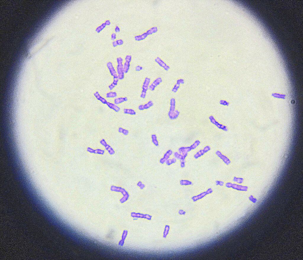
This image is the normal male karyotype. At first glance you may wonder that it’s a mess, But trust me it is one of the best GTG-banding images and it is very difficult to achieve it every time. The image was exclusively taken directly from the microscope with the high quality camera and you may observe some of the very good quality bands on different chromosomes.
Now, this example is of totally normal person. and doesn’t have any abnormalities, care fully observe the beauty of banding pattern. Also, we can count all 46 number of chromosomes.
Examples 2:
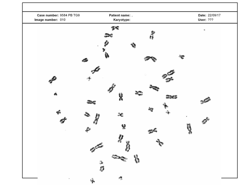
All the examples what i am not explaining are the images taken by the automated microscopy system and given results by the software. See the above example, the banding is not good but chromosomes are separated well enough to count.
Examples:3
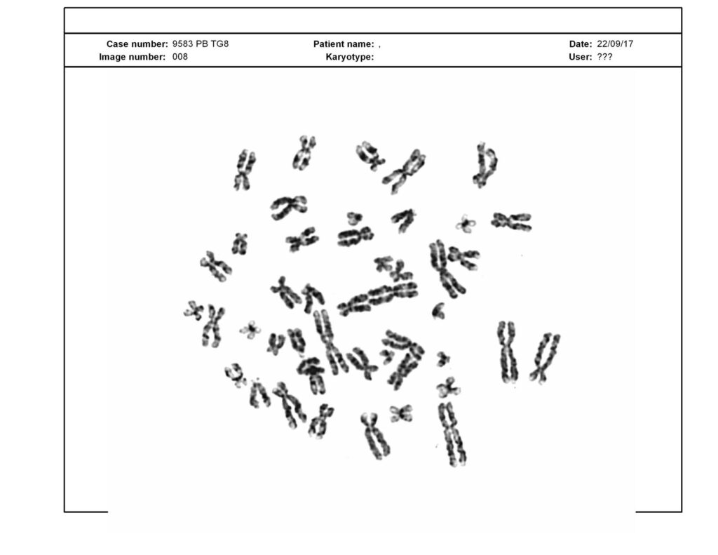
Again the image and banding is not good, but you have observe the chromatins of different chromosomes. You can observe 4 different chromatids clearly. Probably this karyotype is of female (46, XX) because it is not good enough to analyse results.
Example: 4
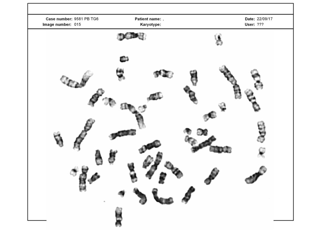
Again, the computer generated results of a karyotype field. the banding patterns are good, the separation and banding pattern is excellent to interpret results. You can count the chromosomes, easily.
Example 5:
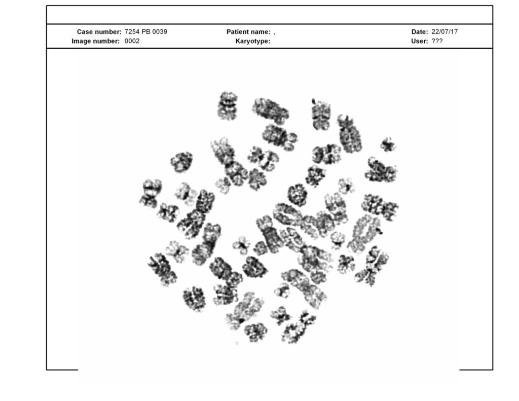
See the example carefully, the field can’t be consider to conclude results because major portion of chromosomes are over digested with trypsin. Besides this, the field is not good enough and chromosomes are not separated well.
Example 6:
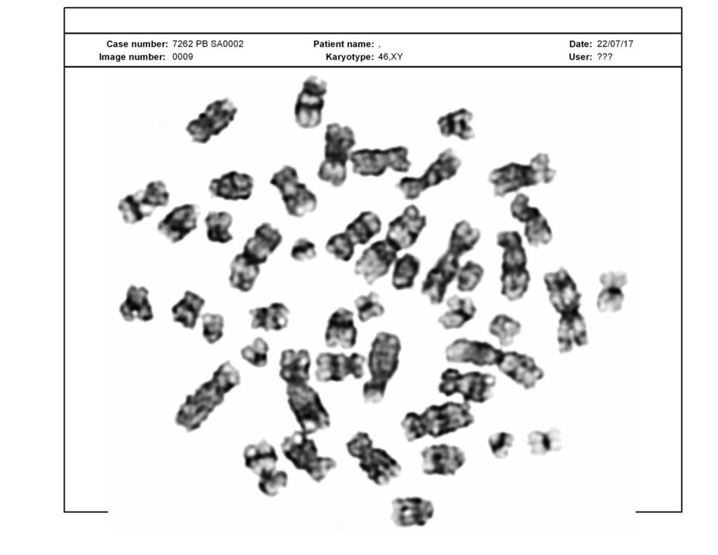
Not a good image to give results, these kinds of fields are usually avoided during analysis. However, you can still observe some good bands on chromosomes. The major portion of chromosomes is digested by the trypsin and that is why less and unclear banding pattern observe.
Next article: What is a karyotype test?
What to observe in a karyotype?
So for a novice, it is a big question, how to interpret or observe a karyotype? But let me tell you first what you can’t observe! in a normal microscope, you can neither observe some of the minor copy number variations like deletions, duplications, nor translocations.
Nonetheless, an expert can do it but for that, he or she too needs to arrange it properly. As a beginner, you can count it directly from the field or by labeling the photograph.
Chromosome numbers one and two are the largest one and metacentric, those 4 you can predict, don’t worry about distinguishing them, we will learn it later.
In the next step, try to find out the acrocentric chromosomes which are 21, 22 and Y and it is easy to find. Collectively, you can now identify chromosome 1, 2, 21, 22, Y and the total number and that is a good start for a newbie.
In addition to this, the X chromosome is small metacentric and can be identified easily. See the image below, i have labelled the above indications for you.
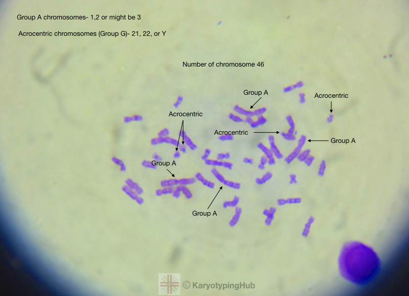
To arrange every chromosome precisely you need to understand the banding pattern of every chromosome and that we will learn in some other article.
We are planning to prepare some assignments for our readers so that they can practice themself. We are also planning to prepare an ebook for you people focusing only on karyotyping only.
Till then, pick one of the examples I have explained, and try to locate the acrocentric chromosomes. the labeled image me on [email protected]. We will feature it in our upcoming article with your name.
Besides all these, to learn how a karyotype is useful and what some common indications and applications of it, please read our previous article: What is a karyotype test used for?
Conclusion:
I hope this article helped you. Those results are our own and from our lab. To give a good experience and how the automated system generates results, I have not edited the image. You can see the patient ID and type of karyogram and image number generated by the system.
I am planning to add some more examples of GTG banding, quinacrine banding and NOR banding image. What you guys say please comment below.

