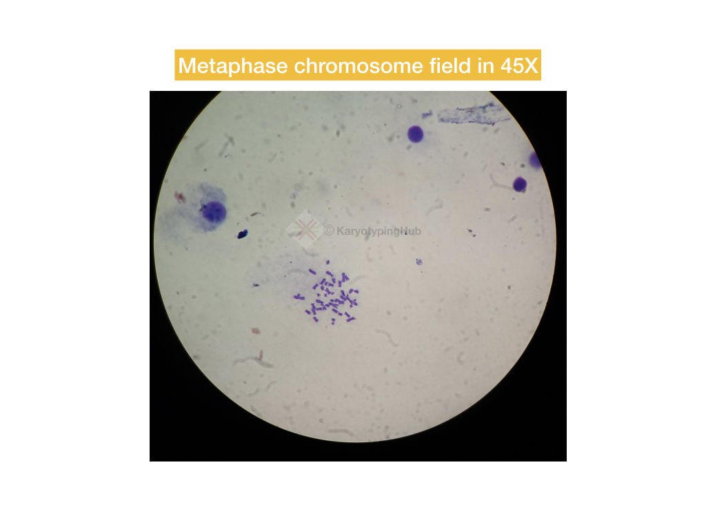
Karyotyping is a process of seeing chromosomes in order to know abnormalities related to it. The metaphase chromosomes are most separatable and visible.
Abnormalities in DNA or chromosomes can cause severe mental, physical or developmental disabilities. In order to find out the alteration related to chromosomes, we have to perform the cytogenetic analysis while to get information of DNA we are performing the molecular genetic tests.
Karyotyping is a common cytogenetic technique or practice, scientists are using it for long. It’s a combination of cell culture, microscopy and genetic analysis and hence a bit complicated.
To perform a karyotype test only a protocol is not enough! We need to have some expertise and experience.
The process of karyotyping initiated by culturing cells in vitro and arresting cells using mitogens.
Not all the cell division phases are karyotyped. A stage at which the chromosomes are the most separate and are used to karyotype. In this article, we will learn which phase or stage of the cell cycle is best for karyotyping and why not others?
Read more: What is Karyotyping?- Definition, Steps, Process, and Advantages.
Which cell division phase is best for karyotyping?
A metaphase stage of mitotic cell division is best to prepare and analyze karyotype. To understand why only the metaphase stage is best! We have to first know the hierarchy of chromosomes.
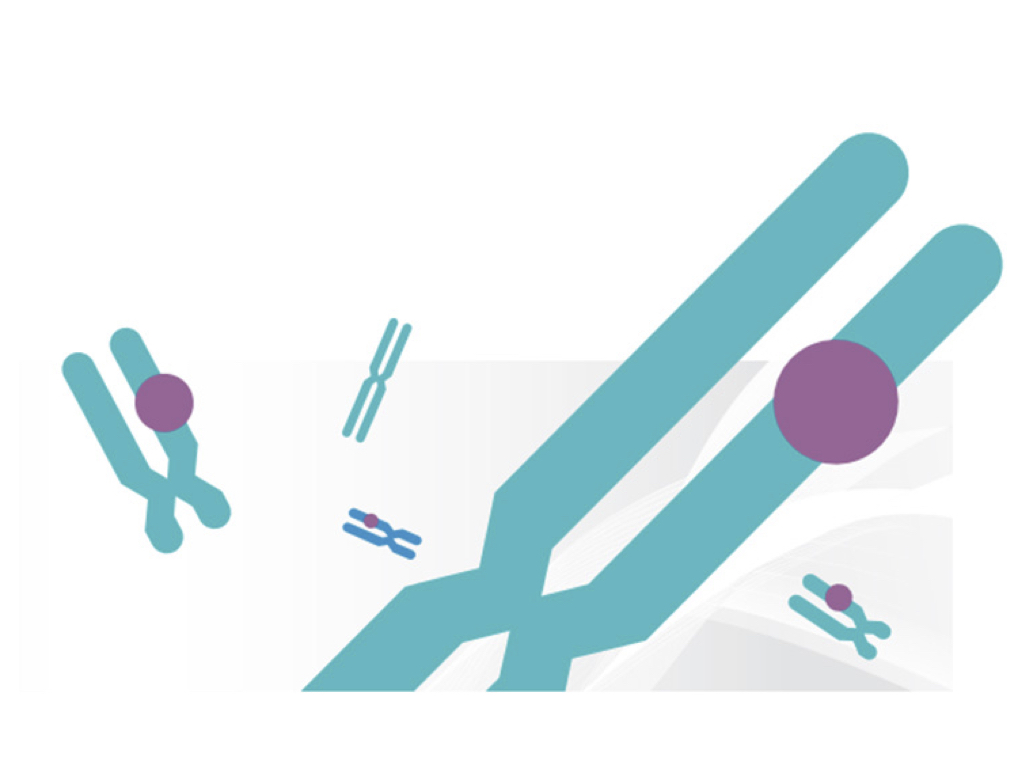
The chromosome is a complex network of DNA and protein which facilitate DNA packaging in a cell.
Usually, chromosomes in other cell division phase aren’t visible or distinguishable because it’s in a relaxed form. The relaxed chromosomes are more released and involved in transcription and translation. But that is not the case with the metaphase.
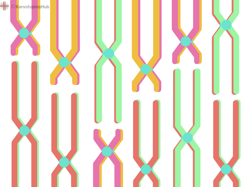
During the metaphase of mitosis, the cells divide and genetic material on chromosomes got separated. The metaphase chromosomes are most condensed and arranged centrally thus are more distinguishable in the microscope.
This is the reason we are using the metaphase stage of cell division to make a karyotype. For that, we are using the PHA-P- a type of mitogen that arrests cells on metaphase and induces rapid cell division.
In order to make it more separatable, we also use colchicine.
Interestingly, sometimes interphase chromosomes are also targeted for karyotyping to study the sister chromatin exchange.
Related article: Colchicine- A Mitotic Inhibitor in Karyotyping.
DNA during a different phase of cell division:
Note that it is just a brief overview of the cell cycle not explained in detail.
Prophase:
The prophase is the stage of initiation of cell division in which the DNA starts making chromosomes by interaction with the proteins. Also, the process of nuclear membrane breakdown starts and the centrosomes start moving to the poles.
Meanwhile, spindle fibers appear too.
Metaphase:
Chromosome packed tightly and lineup at the metaphase plate. The spindle fibers start attaching with the centromeres. Each sister chromatids attached to the spindle fibers.

Anaphase:
The centromere and slit into parts and moves to the opposite pole along with the sister chromatids which are now become whole chromosomes for a new cell.
Telophase and cytokinesis:
The spindle fibers break and chromosomes are moved to two daughter cells. The nuclear membrane forms and two daughter cells with the same number of chromosomes are formed.
Conclusion:
The metaphase is the best phase or stage with separate chromosomes and exactly the diploid one. So that we can identify and arrange its chromosome properly. All genetic material of a cell is arranged on a metaphase chromosome and makes euchromatin and heterochromatin regions.
The euchromatin and heterochromatin regions are stained differently and make different banding patterns that are why the metaphase chromosomes are widely used in karyotyping.


