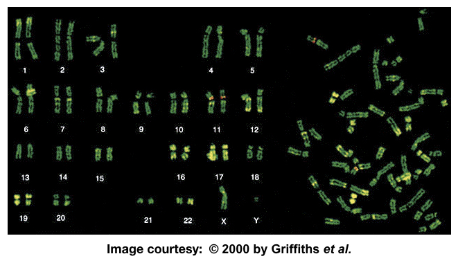
“The Q banding or quinacrine banding technique in which the fluorescence dye quinacrine binds or intercalates with the DNA and gives different banding patterns on chromosomes.”
The technique is also known as QFQ banding- Q-bands using fluorescence quinacrine and was first explained by Caspersson and co-workers.
Related article: 20 Common karyotyping (or Chromosomal) Abnormalities.
What is Q banding?
The Q banding is a Quinacribe banding based on the use of fluorophore quinacrine or atebrin. It’s a type of intercalating dye that binds to DNA and emits fluorescence when observed under a microscope.
Therefore, to observe chromosome bands using the present technique, a fluorescent microscope is required.
Much like the G bands the process of Q banding is simple, effective, rapid and doesn’t require pre-aged slides. Heating and incubation steps aren’t needed too.
Old, aged or freshly prepared slides can be processed using the present banding technique, however, microscopy must be performed immediately as soon as possible.
Differential banding patterns observed on different chromosomes note that the Y chromosomes show a huge bright fluorescence and are most intensely stained.
Pro-tips: The brightest chromosome in the QFQ bands is the Y chromosome.
Principle:
When stained with the quinacrine, the AT regions enhance the fluorescence while the GC rich regions quench. More this, the base composition of DNA, the length of the chromosome and protein DNA interaction plays an important role in banding patterns of QFQ banding.

Requirements:
Slides and coverslips, Fluorescence microscope, Quinacrine dye, Quinacrine dihydrochloride solution, coupling jar, distilled water, MacIIvain’s buffer.
Related document: Utilities for karyotyping.
Protocol:
- Place the slides in a Coplin jar having quinacrine dihydrochloride solution to stain for few minutes.
- Rinse slides in tap water or distilled water to remove excess stain and put an ultra-thin coverslip.
- Rinse it with tissue paper and immediately observed under the fluorescence microscope. If possible seal slides with rubber cement.
- Capture images as soon as possible.
Caution: Inhaling, swallowing and skin contact of quinacrine dihydrochloride is harmful and not advisable.
Key points:
Expert hands are required to investigate QFQ banded chromosomes. Mishandling of fluorescence microscope and poor illumination is the common reason for weak banding or fluorescence. This can happen because of inappropriate chromosome treatment with the Quinacrine.
Chromosome fading occurs due to excessive buffer on the slide.
High fluorescence may be observed due to incorrect immersion oil or quinacrine treatment for a longer time.
Application:
Much like the GTG banding, the QFQ banding is widely used in investigating various chromosomal abnormalities because of their distinct and uniform banding pattern.
Heteromorphisms of chromosomes 3, 4 and Y can be studied more accurately and can be used as a marker to study various chromosomal aberrations.
Conclusion:
Quinacrine banding was developed first and widely used for a long time. However, the use of fluorochromes makes it costlier and not so common for banding. Researchers and cytogeneticist use GTG banding more commonly in routine experiments.
Source:
Human chromosomes: Principle and Techniques (Second Edition) by Ram S. Verma and Arvind Babu.


