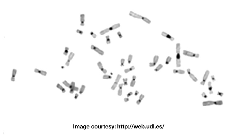
“The C banding or C bands is a technique utilized in karyotyping which stains especially the centromeric region, stronger than other regions.”
Do you know?
- The centromere region of chromosomes is a repetitive DNA region, condensed and the non-active region.
- It distinguished two arms from each other.
- Centromere helps in the separation of chromosomes or sister chromatids during cell division.
- 46 chromosomes mean 46 centromeres are there in a genome.
- It helps to hold arms.
Telomere, centromere and chromosome arms are three distinct parts of chromosomes in which centromere and telomeres are repetitive DNA-rich regions.
The centromere actually helps in the segregation or separation of chromosomes during cell division which facilitates replication afterward.
A specialized banding technique is known as centromere banding or C bands or C banding is used to study the centromeric region of chromosomes.
Related article: What is Chromosomal Translocation?- Definition, Mechanism and Types.
What is a C banding?
The C bands stain the centromeric constitutive heterochromatin regions of a chromosome. This portion contains mostly repetitive DNA, satellite DNA and a little non-repetitive/ non-coding DNA which are present during the interphase stage of mitosis.
The constitutive heterochromatin regions comprising satellite DNA are mostly AT-rich regions.
The constitutive heterochromatin region is non-coding regions which are gene-lacks. The original technique was explained by Arrighi and Hsu in 1971 using alkali and sodium hydroxide preliminarily.
However, in 1972, Sumner explained the improvised version of the present technique using mild alkaline and Barium hydroxide instead of sodium hydroxide.
C bands stain the center region, the centromere of chromosomes, note that on the Y chromosome, it is observed on the distal region of the long q arm.
Principle of C banding:
In order to stain only the centromeric portion, we need to denature or break all the chromosomal DNA, besides the constitutive heterochromatin.
Treatment of alkaline reagent and acid (HCl) denatures DNA and breakdown the non-constitutive heterochromatin DNA. In successive washing steps, the breakdown DNA from chromosome arms is removed but C bands remain intact. Though the clear reason why the DNA present on the constitutive heterochromatin regions remains untreated isn’t yet clear. It is believed that some special histone proteins which are only present near the C bands prevent denaturation.
Note that the clarity of bands depends on the concentration of chemicals, temperature and duration.
Read more: What is Karyotyping?- Definition, Steps, Process, and Advantages.
Requirements:
Pre-aged slides, coupling jars, Barium hydroxide, sodium hydroxide, HCl, 2X SSC solution, Giemsa stain, phosphate buffer, water bath and coverslips.

Protocol:
C bands using NaOH:
- Treat pre-aged slides in HCl solution for few minutes at room temperature and was slides in either phosphate buffer or distilled water.
- Apply a few drops of RNase solution on slides, cover it coverslip and incubate it for an hour at room temperature.
- Place slides in NaOH solution for a few minutes at room temperature.
- Immediately wash slides in various combinations of alcohols to remove the NaOH as soon as possible.
- Allow slides to heat in pre-heated coupling jars at 65°C in SSC buffer overnight.
- The next day, wash slides using distilled water and air dry it.
- Stains slide in 5% Giemsa solution in Sorensen’s buffer (pH 7.0) at room temperature for few minutes.
- Observed slides in bright field or phase-contrast microscope.
You can use barium hydroxide instead of sodium hydroxide, using the same protocol, although the concentration may vary. Note that instead of overnight allow slides to heat in the SSC for 2 hours at 65°C.
C Bands using Barium hydroxide (Ba(OH)2):
The protocol of using Barium hydroxide is a bit different than the sodium hydroxide, the reason is the strong reactivity of barium hydroxide to atmospheric carbon dioxide.
While performing C banding using barium hydroxide, every time prepare the Ba(OH)2 solution freshly, filter it and store it in a closed container. Also, fash slides thoroughly to avoid the deposition of barium hydroxide crystals.
In comparison to the NaOH, the barium hydroxide is milder alkali and therefore reacts slowly. So instead of 60C, perform C bands using the present technique at room temperature which preserves the morphology of chromosomes.
Either of the reagents performs denaturation of DNA and henceforth treatment for a longer time destroys the morphology of chromosomes while shorter duration treatments can’t provide interpretable C bands.
Experts need to decide the dose or concentration of either chemical and duration depending upon the age of slides.
Overnight treatment and washing play important roles in results development.
Here is the brief protocol of C bands using barium hydroxide:
- Treat pre-aged slides in HCl solution for few minutes at room temperature and was slides in either phosphate buffer or distilled water.
- Apply a few drops of RNase solution on slides, cover it coverslip and incubate it for an hour at room temperature (optional).
- Place slides in Ba(OH)2 solution for 30 minutes at room temperature or 10 minutes at 50 to 60°C temperature.
- Immediately wash slides in various combinations of alcohols to remove the NaOH as soon as possible.
- Allow slides to heat in pre-heated coupling jars at 65°C in SSC for 2 hours.
- Wash slides using distilled water and air dry them.
- Stains slide in 5% Giemsa solution in Sorensen’s buffer (pH 7.0) at room temperature for few minutes.
- Observed slides in bright field or phase-contrast microscope.
To run the protocol of C bands few points should be taken into consideration while performing the protocol. Here they are:
Use only the pre-aged slides. Freshly prepared slides are too sensitive and can’t handle harsh chemical treatments, hence it damages the morphology.
To do so prepare slides, air dry them and store them at cool or dry places for a week. Or
Age slides for 60°C temperature for 2 to 3 days.
Another important point is a combination of chemicals and treatment.
Every lab or cytogenetic lab has a different environment and conditions therefore, the concentration of chemicals and duration of treatment may differ.
No such exact protocol can work in this case, therefore, prepare different replicates having different concentrations of either NaOH or Ba(OH)2.
Prepare at least 3 to 6 sets of reactions to get good results. Once you get results use the same formula or recipe next time.
RNase step can be avoided as it hasn’t directly affected the C bands or constitutive heterochromatin regions.
Caution: Barium hydroxide is highly toxic and corrosive, use under the supervision of experts.
Applications:
The technique is used to study chromosome rearrangements, especially at the centromeric region of chromosomes.
Translocation in which the centromeres are involved can be studied effectively using the present technique.
C bands polymorphism:
The C bands of all chromosomes in all individuals are polymorphically known as C bands hetero-polymorphism or C band polymorphism. The C bands of different chromosomes in an individual or between individuals vary greatly.
Based on their size, categorized into 5 different categories which are used to study various polymorphisms.
It is useful in studying the chromosomes of unusual morphology. However, the C bands polymorphism do not have clinical significance but are inherited familial.
Read further: 10 best karyotyping activities and assignments.
Conclusion:
As we said, the C banding stains the centromeric region of chromosomes, however, the C bands of chromosomes Y, 1, 9 and 16 have a larger C band than the other.
The present technique is more preferred when studying the copy number variations near the constitutive heterochromatin regions. Note that it can’t stain other regions of chromosomes. No banding patterns are observed on chromosome arms.
Source:
Human chromosomes: Principle and Techniques (Second Edition) by Ram S. Verma and Arvind Babu.


