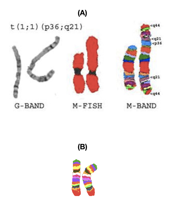
M-Bands or M Banding is a high-resolution chromosome banding technique that finds cryptic chromosomal rearrangements such as inversion, translocation, deletion or duplication at the chromosome level.
Banding techniques have a significant role in cytogenetics especially in karyotyping. The whole cytogenetic studies are conducted around chromosomes and to encounter chromosomal anomalies. Many techniques and improvements over a period of time facilitated accuracy in the investigation.
Karyotyping is a true, conventional cytogenetic technique that finds various chromosomal abnormalities and utilities since years. Although recent advancements like multicolor FISH and spectral karyotyping are far more accurate and wide range of investigation advantages over karyotyping.
Nonetheless, conventional karyotyping using the GTG banding is a cost-effective, reliable and most trusted method in evaluating numerical chromosomal abnormalities but not for copy number variations.
To conduct various chromosomal studies different banding techniques like G bands, C bands, Q bands, NOR bands, T bands and high resolution banding are utilities in cytogenetic labs.
Interestingly, the mbands or m banding isn’t a conventional banding technique, evolved in recent years but used to investigate smaller chromosomal variations.
Comprehensively if we describe banding patterns or how it works? Banding stains different chromosome regions and provides varied dark or light band patterns which different from chromosome to chromosome. Ultimately, it makes chromosomes distinguishable from each other.
Through present content, we will explain to you one of the most advanced and state of the art banding techniques the “M- bands” or multicolor banding.
What is M banding?
The m banding is also known as mBands, multicolor bands, m chromosomal banding or multicolour chromosomal banding technique stains different regions of a single chromosome with different colors.
It’s fluorescent chemistry based technique needs a fluorescent microscope and various color filters to investigate results. It has a few megabase pair range of resolution which is slightly lower than the conventional one! But different colored bands on a single chromosome facilitate ease in investigation.
Requirements:
Cell culture- sodium heparin tubes, blood sample, l-glutamine, cell culture media- RPMI 1640, phytohemagglutinin, colcemid, methanol, glacial acetic acid, slides, G stain.
M banding- ready to use m banding probes, alkaline reagent, hybridization kit, metaphase slides, fluorescent microscope, analysis software.
Other utilities- CO2 incubator, safety cabinet, centrifuge, compound microscope, GTG banding chemicals, hypotonic solution and other requirements for karyotyping.
Related article: Comparison Between Karyotyping and Microarray.
Principle of M-banding:
Probes prepared by multiple chromosome region-specific DNA libraries and labeled with different colored fluorochromes are applied to hybridize with chromosome regions.
Through alkaline denaturation, probes bound to different chromosome regions emit fluorescence under fluorescence microscope. A system software creates images of chromosomes based on the signal obtained.
Results show multicolor bands on the chromosome regions that are distinguishable.
Process of M-Banding:
M banding or multicolor chromosome banding needs metaphase chromosomes to study. Cells are cultured as per standard karyotyping protocol and harvested. Slides are prepared as indicated in the karyotyping protocol and placed to age.
The m banding probes which are different from the mFISH probes are synthesised using the pooled multiple chromosome region specific DNA libraries.
Synthesised probes usually are 20 to 40 nucleotides long, labelled with fluorochromes.
Alkaline denaturation is performed in order to digest or denature chromosomal DNA regions followed by probe hybridization.
Incubate slides overnight to hybridize probes effectively and wash it with a buffer (provided by manufacturer) or distilled water.
Wet slides for microscopic analysis.
An image analysis software prepares the coloured image of chromosomes based on the fluorescence emitted by various probes hybridised.
Differential multicolor banding patterns help to investigate and interpret results of karyotyping.
Note that the m banding technique and mFISH technique are different.
M banding vs M-FISH:
The mFISH is a variation of fluorescence in situ hybridization in which each chromosome or chromosome arms are colored using a different set of fluorescent probes.
The m banding technique is a variation of standard karyotyping in which a single chromosome is colored with a different set of probes.
mFISH is often known as spectral karyotyping, different sets of chromosomes or chromosome arms are hybridized with different probes.
The m banding uses different colored probes to make a differential banding pattern or different colored banding pattern on a single or pair of chromosomes.
mFISH investigates translocation or deletion effectively whilst m banding identifies interchromosomal exchanges, deletons, inversions or translocations.
However, both techniques depend on fluorescent chemistry, utilising probes of different colors and processed through alkaline denaturation followed by hybridization.
Advantages of M- banding:
There are a couple of excellent advantages of m banding over conventional GTG bands.
- On single chromosomes varied colored bands can be seen.
- Bands are well separated and distinguishable from one another.
- Abnormalities can be mapped correctly.
- More accurate and effective than conventional karyotyping.
Applications of M- banding:
- The technique finds cryptic chromosomal abnormalities.
- Scientists use m banding in cancer studies and finding markers associated with a particular type of cancer.
- It can find interchromosomal deletions, duplications, insertions or translocations.
- It can investigate complex chromosome rearrangements.
Limitations:
- PCPs- partial chromosome paint probes specific to m banding sometimes overlap on chromosome regions cause a decrease in fluorescence intensity, create ambiguity in results, consequently.
- Can’t finds rearrangements, translocations or copy number variations between two chromosomes.
- Can’t identify minor copy number variations.
- It’s a low-resolution technique.
- Needs fluorescence set up to investigate results.
In recent times, different probes or probe sets are available to investigate different chromosomal rearrangements.
Other banding techniques: Q-Banding, C-Banding, T-Banding.
Comparison of techniques:
| Technique | Applications |
| Conventional karyotyping | Numerical chromosomal abnormalities, larger deletions or duplication. |
| M banding | Interchromosomal rearrangements, duplications, deletion, insertion or translocations. |
| FISH | Complex chromosomal rearrangements and copy number variations. |
| SKY | Differentiating chromosomes, chromosome regions, translocation and duplications between chromosomes and other complex rearrangements. |
| Array CGH | Minor copy number variants which can’t be investigated using FISH or SKY. Many Thousands of chromosomal rearrangements and copy number variants at once. |
Conclusion:
Molecular cytogenetic techniques like FISH, SKY and array CGH evolved recently and are more advantageous than the conventional karyotyping. SKY- spectral karyotyping or FISh- fluorescent in situ hybridization finds breaks and rearrangements between more than two chromosomes which can’t be identified by conventional technique.
However, those even can’t help to investigate uni-chromosomal rearrangements or abnormalities. M banding helps to investigate and identify those rearrangements, deletions, duplication or translocations occurring on a single chromosome. Note that it has lower banding resolution.
Sources:
Hu J, Sathanoori M, Kochmar SJ, Surti U. Application of multicolor banding for identification of complex chromosome 18 rearrangements. J Mol Diagn. 2006;8(4):521-528. doi:10.2353/jmoldx.2006.060001