A karyotype report is a written proof of what a cytogeneticist observes from your sample.
Usually, medical reports, especially the genetic reports like the karyotype or real-time PCR report are complicated to understand, even for a doctor.
Report is actually a written proof about your testing, and the investigator takes responsibility for it. However, as per the standard rules, the medical reports including the karyotype report should be self explanatory to understand.
To perform, evaluate and understand testing techniques like karyotyping needs high end expertise and experience. It’s more manual than automated, everything depends on the expert hands, spart eyesight and previous experience.
Performing understanding and evaluating karyotyping like technique is actually a difficult task, indeed, geneticists tried to make a self explanatory report format.
In the present article, I will explain to you the standard format of karyotyping reporting, what should be there and what not. Also what the results indicate.
Karyotyping report:
The reporting format can be broadly divided into three or four sections; General or primary information, technical information, graphs and images of results, if any and other information.
Firstly the karyotyping report or any other reporting format should have servera basic informations in it. Information that shows who the patient is, what the sample type and when the sample is received.
Also it should show the sample type in it. The reason is, different combinations of chemicals and culture media composition are needed for different sample types.
Related reads:
Primitive informations:
- Name of the patient
- Guardian’s name
- Address of the patient
- Contact detail
- Doctor’s name and address who referred the patient
- The date of sample collection
- Date of sample received
- Date of sample processed
- The type of sample received
- The condition of sample when received
These primary information must be there on every medical testing report, on the front page. As we said, the report is self explanatory, anyone can know the status of sample and patient and hence above listed information must be on the front as well as on the top of the page.
Technical informations:
Afterward, the next section should have technical information in it like what technique is employed, how it is used, overview of technique used etc etc.
Again the reason to use this type of information is to make an expert or doctor understand the condition of testing.
Technical information:
- Technique employed
- Criteria of testing
- Condition of testing
- Range of error of testing technique
- Limitations of instrument used in the testing
Note that this information is used during the non-redundancy of the results so that the expert can understand under which conditions the test was performed.
Graphs, image or pictures:
The next section is included in the reporting format to make the report impressive and look attractive, though the image, graph and picture of the results are valuable evidence in some critical medical testing.
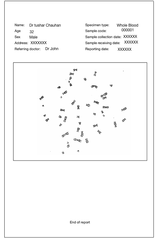
For example, the Ct value graph must be included in the RT PCR report. Images of the fetus must be included in the sonography report and pictures of field and observations must be there in the karyotyping report.
Again, the reason behind including this section is to make the expert understand the results. Some doctors or scientists are expert enough to understand the results even by only seeing the picture or graph therefore it is there in the report.
Moreover, it is a proof about what is there in your sample.
Other information:
Based on the type of report and requirement of the reporter other information is included in the reports to understand the patient’s condition more accurately.
For example, If a doctor has sent a sample to study the Philadelphia translocation and the karyotype evaluator observes nothing or has confusion about what to do, he or she can indicate further testing like FISH or microarray.
Therefore a dedicated “indication” or “note” section is included at the end of the report.
These informations are compulsorily needed on the karyotype report or any other medical report however, some minor changes are acceptable. Some testing companies also included normal and abnormal testing ranges and terms and conditions of reporting in their report as well.
Note that a medico-legal disclaimer must include in the back of the report so that a patient can understand in which condition he or she can claim the report.
Example of karyotyping or karyotype report:
Here we have enlisted a standard karyotype report format and one example showing all the information of the patient. To make you understand the report I had given my own example, note that it is just a dummy report.
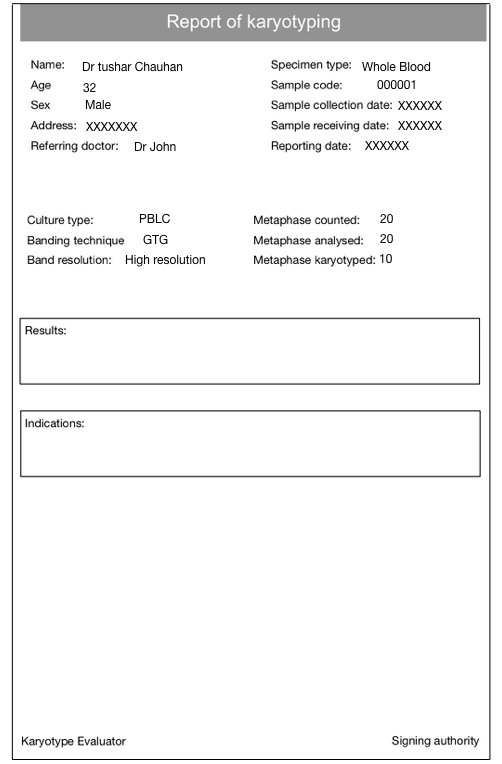
On the top of the report, we have all the information of the patient and sample. Afterward, we have information about karyotyping technique indicating that we have used peripheral blood lymphocyte culture technique to culture blood cells.
We have used G-bands by trypsin and Giemsa technique to band chromosomes.
A high resolution banding approach is employed to study the results.
Other in depth information shows that,
We have counted a total 20 metaphase plates or cells to investigate the sample in which we have observed all 20 metaphase fields.
We have prepared 10 karyotypes from all the 20 metaphase fields and finally given the results shown in the image.
After that, the results are explained as per the ISCN nomenclature. If you want to learn what the ISCN nomenclature is, you can read this article: ISCN nomenclature system for human karyotype.
If any abnormalities are observed, that must be indicated in the picture given in the report.
At the end of the human karyotype report, the section for signing authority and karyotype evaluator is included that increases the authenticity of the report.
The sign of the expert with their name, designation in the lab and degree increases the trust factor of referring doctors as well as patients to many folds.
What should a normal report of karyotyping or cytogenetic shows?
A normal karyotype report show a normal karyogram and some common information related to report. No abnormalities are indicated in the report.
Conclusion:
I had performed karyotype of many patients, signed many reports, and seen various types of reporting formats during my professional career. This reporting format that I had given here is prepared with the best knowledge of mine.
The format may change from lab to lab and researcher to researcher. Though some crucial and ‘must need’ information must be there on every report.