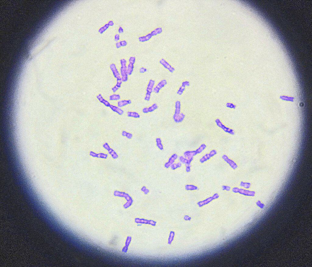
GTG banding also is known as the Giemsa-trypsin-Giemsa banding technique is employed during karyotyping to investigate chromosomes and chromosomal anomalies.
Chromosomal anomalies like structural aberrations or numerical aberrations are commonly encountered using different types of banding techniques. GTG banding, R-banding, NOR-banding and Q- banding are popular techniques used routinely.
However, the GTG banding technique used so commonly to investigate chromosomes. The GTG banding technique is simple and easy to use. Also fluorescence dyes are not utilized in it.
The present technique is a combination of staining and enzymatic activity. In the present article we are going to learn the commonest GTG banding technique.
Interesting articles:
- A karyotype of Down syndrome
- A karyotype of Patau syndrome
- A karyotype of Klinefelter syndrome
- A karyotype of Edward syndrome
Principle:
The GTG banding technique is employed in order to study the chromosomes and investigate chromosomal aberrations. Here the enzyme trypsin digests the protein portion of the chromosomes (some portion for some times), because we are not treating slides for longer times.
Meanwhile, the Giemsa solution stains the chromosome, more condensed portion stain lightly while the relaxed portion stain darky.
The trypsin digest the histones and allow chromatin relaxation. At the same time, treatment of both the agents makes different banding patterns on chromosomes.
Requirement:
Giemsa solution: 5%
- Distilled water: 42.5 ml
- Buffer solution(Gurr’s pH 6.8) : 5.0 ml
- Giemsa stain: 2.5 ml.
- Total: 50 ml
Trypsin solution: (0.05%)
- Trypsin (1: 250): 50 mg
- Dulbecco’s phosphate buffer saline- CMF: 100 ml
Procedure:
Age the slide for 1 before performing the banding for 80°C to 90°C or prepare the air-dry slide overnight for 55°C to 60°C.
Dip th slides in the 0.05% trypsin solution for 5 to 10 minutes (freshly prepared).
Rinse the slide with the PBS and stain it with the Giemsa stain for 4 to 6 minutes.
Rinse the slide with the running tap water and allow the slide to dry.
Observe the slide directly under the microscope without using the coverslip.
Some recommendations:
One of the important things to keep in mind while performing the banding is that prepare the GTG banding solution freshly every time. However, you can use a single batch for a whole day.
Store the trypsin solution at 4C.
Use the trypsin and giemsa solution for 3 to 4 hours. The solution becomes turbid, discard it immediately or if contamination seen on slide immediately discard the solution.
The crucial factor for the GTG banding is the wise use of time. The longer you treat the slide, the more banding will disturb. However, if you treat slides for less time, the banding pattern will not be developed correctly. Hence in order to make a distinguishable banding pattern, you have to optimize the protocol by yourself.
For that use a different combination of time and observe slides. Choose the time for treatment that gives you good results.
Alterations:
In order to optimize the accuracy of results, different combinations of Giemsa and trypsin with different combinations of time are used. Here some are;
0.5% trypsin (10-60s) and 10% giemsa (8 min – 15 min)
- 1% trypsin (10-60s) and 10% Giemsa (8 min – 15 min)
- using 0.5% trypsin (10-60s) and 5% Giemsa (8 min – 15 min)
- 1% trypsin (10-60s) and 5% Giemsa (8 min – 15 min)
- Allow the slide to rest in the refrigeration 4°C for a while to stop the activity of trypsin.
Start to read from here: What is Karyotyping?- Definition, Steps, Process, and Advantages.
Conclusion:
The present GTG banding protocol is good enough to study chromosomes, though, additional optimizations are needed to master the technique. Remember, If you treat the slide in trypsin for longer time, all the proteins will digested and banding will not be appear.
Source:
Human chromosomes: Principle and Techniques (Second Edition) by Ram S. Verma and Arvind Babu.